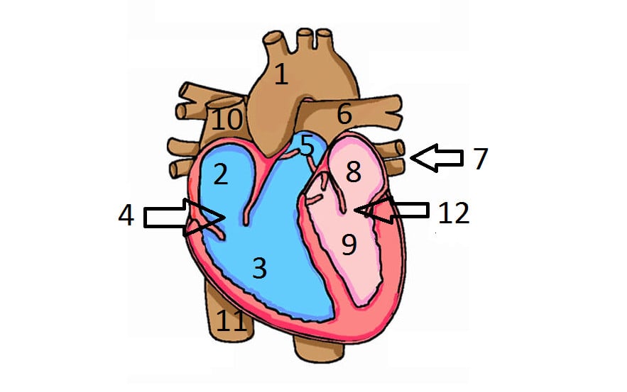
42 the human heart and its labels
File:Diagram of the human heart (no labels).svg - Wikimedia This file is licensed under the Creative Commons Attribution-Share Alike 4.0 International license.: You are free: to share - to copy, distribute and transmit the work; to remix - to adapt the work; Under the following conditions: attribution - You must give appropriate credit, provide a link to the license, and indicate if changes were made. You may do so in any reasonable manner, but ... Heart: illustrated anatomy - e-Anatomy - IMAIOS This interactive atlas of human heart anatomy is based on medical illustrations and cadaver photography. The user can show or hide the anatomical labels which provide a useful tool to create illustrations perfectly adapted for teaching. Anatomy of the heart: anatomical illustrations and structures, 3D model and photographs of dissection.
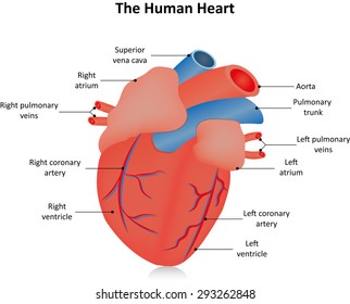
PDF Anatomy of Heart Labeled and Unlabeled Images aortic arch left pulmonary artery left pulmonary veins auricle of left atrium left atrium circumflex artery (in atrioventricular sulcus) coronary sinus left ventricle (c) posterior view of the external heart © 2019 pearson education, inc. ascending aorta superior vena cava right pulmonary artery right pulmonary veins right atrium inferior vena …

The human heart and its labels
bodytomy.com › labeled-diagram-of-human-heartA Labeled Diagram of the Human Heart You Really Need to See The human heart, comprises four chambers: right atrium, left atrium, right ventricle and left ventricle. The two upper chambers are called the left and the right atria, and the two lower chambers are known as the left and the right ventricles. The two atria and ventricles are separated from each other by a muscle wall called 'septum'. › heart › picture-of-the-heartHuman Heart (Anatomy): Diagram, Function, Chambers, Location ... The heart is a muscular organ about the size of a fist, located just behind and slightly left of the breastbone. The heart pumps blood through the network of arteries and veins called the ... › Heart-Diagram-LabeledDiagram of Human Heart and Blood Circulation in It | New ... Exterior of the Human Heart A heart diagram labeled will provide plenty of information about the structure of your heart, including the wall of your heart. The wall of the heart has three different layers, such as the Myocardium, the Epicardium, and the Endocardium. Here's more about these three layers. Epicardium
The human heart and its labels. byjus.com › biology › diagram-of-heartHeart Diagram with Labels and Detailed Explanation - BYJUS Well-Labelled Diagram of Heart. The heart is made up of four chambers: The upper two chambers of the heart are called auricles. The lower two chambers of the heart are called ventricles. The heart wall is made up of three layers: The outer layer of the heart wall is called epicardium. The middle layer of the heart wall is called myocardium. The inner layer of the heart wall is called endocardium. Heart Labeling Quiz: How Much You Know About Heart Labeling? Here is a Heart labeling quiz for you. The human heart is a vital organ for every human. The more healthy your heart is, the longer the chances you have of surviving, so you better take care of it. Take the following quiz to know how much you know about your heart. Questions and Answers. 1. Human Heart-Label Diagram | Quizlet Label the diagram Learn with flashcards, games, and more — for free. Home Browse. Browse. Languages. English French German Latin Spanish View all. Science. Biology Chemistry Earth Science Physics Space Science View all. Arts and Humanities. ... Human Heart-Label. STUDY. Learn. Write. Test. PLAY. How to Draw the Internal Structure of the Heart - wikiHow Make sure to label the following: Superior Vena Cava Inferior Vena Cava Pulmonary Artery Pulmonary Veins Left Ventricle Right Ventricle Left Atrium Right Atrium Mitral Valves Aortic Valves Aorta Pulmonic Valve (Optional) Tricuspid Valve (Optional) 6 To finish, label "The Human Heart" above the sketch. Community Q&A Search Add New Question Question
Human heart label Images, Stock Photos & Vectors | Shutterstock Find Human heart label stock images in HD and millions of other royalty-free stock photos, illustrations and vectors in the Shutterstock collection. Thousands of new, high-quality pictures added every day. Heart Diagram with Labels and Detailed Explanation The heart is located under the ribcage, between the lungs and above the diaphragm. It weighs about 10.5 ounces and is cone shaped in structure. It consists of the following parts: Heart Detailed Diagram Heart - Chambers There are four chambers of the heart . The upper two chambers are the auricles and the lower two are called ventricles. How to Draw a Human Heart: 11 Steps (with Pictures) - wikiHow If you're trying to identify parts of the heart for a class you're taking, it's good practice to draw the heart yourself and label each segment. You can refer to your textbook in order to label the: Aorta Superior vena cava Inferior vena cava Right and left atria Right and left ventricles Pulmonary veins and arteries 5 Heart Anatomy: Labeled Diagram, Structures, Blood Flow ... - EZmed Image: Use the 2x2 table to label the 4 chambers of the heart, including the right atrium, right ventricle, left atrium, and left ventricle. Tricuspid Valve and Mitral Valve Now that we have a good understanding of the 4 chambers of the heart, let's move on to the 4 main valves.
sciencetrends.com › human-heart-diagram-labeledHuman Heart Diagram Labeled | Science Trends Human Heart Diagram Labeled Daniel Nelson 1, January 2019 | Last Updated: 3, March 2020 The human heart is an organ responsible for pumping blood through the body, moving the blood (which carries valuable oxygen) to all the tissues in the body. Without the heart, the tissues couldn't get the oxygen they need and would die. Anatomy of a Human Heart - uofmhealth Located between the lungs in the middle of the chest, the heart pumps blood through the network of arteries and veins known as the cardiovascular system. It pushes blood to the body's organs, tissues and cells. Blood delivers oxygen and nutrients to every cell and removes the carbon dioxide and other waste products made by those cells. Human Heart - Diagram and Anatomy of the Heart - Innerbody The heart is a muscular organ about the size of a closed fist that functions as the body's circulatory pump. It takes in deoxygenated blood through the veins and delivers it to the lungs for oxygenation before pumping it into the various arteries (which provide oxygen and nutrients to body tissues by transporting the blood throughout the body). Label the heart — Science Learning Hub Label the heart Add to collection In this interactive, you can label parts of the human heart. Drag and drop the text labels onto the boxes next to the diagram. Selecting or hovering over a box will highlight each area in the diagram. Pulmonary vein Right atrium Semilunar valve Left ventricle Vena cava Right ventricle Pulmonary artery Aorta
Biology Diagrams,Images,Pictures of Human anatomy and physiology : Exchange of gases in lungs ...
Heart: Anatomy and Function Your heart is the main organ of your cardiovascular system, a network of blood vessels that pumps blood throughout your body. It also works with other body systems to control your heart rate and blood pressure. Your family history, personal health history and lifestyle all affect how well your heart works. Appointments 800.659.7822
.png/120px-Diagram_of_the_human_heart_(catalan).png)
File:Diagram of the human heart (cropped).svg - Wikimedia Commons
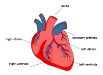
The structure of the heart - Structure and function of the heart ... - BBC The heart is a large muscular pump and is divided into two halves - the right-hand side and the left-hand side. The right-hand side of the heart is responsible for pumping deoxygenated blood to ...

Brain - Examville
Label the Human Heart - 6th Grade Science Worksheets - SoD Label the Human Heart. Your heart is one of the most vital organs in your body. It's a pump that works round the clock to ensure the constant flow of blood throughout the circulatory system. Now it's time to show how much you know about your own heart. Get started by labeling its different parts with this cool science worksheet for 6th grade.

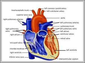
A Diagram of the Heart and Its Functioning Explained in Detail The heart blood flow diagram (flowchart) given below will help you to understand the pathway of blood through the heart.Initial five points denotes impure or deoxygenated blood and the last five points denotes pure or oxygenated blood. 1.Different Parts of the Body ↓ 2.Major Veins ↓ 3.Right Atrium ↓ 4.Right Ventricle ↓ 5.Pulmonary Artery ↓ 6.Lungs

DRAW IT NEAT : How to draw human baby in womb
Heart Labeled Stock Illustrations - 213 Heart Labeled ... - Dreamstime Sketch of human heart anatomy on blue line on a white background. Educational diagram showing blood flow with hand written labels of the main parts. Vector. Wooden wall labeled - I love my boss. Colored wooden wall labeled - I love my boss, 3d render. Graves disease vector illustration. Labeled diagnosis symptoms diagram.

Human Heart Labeling Worksheets & Teaching Resources | TpT Human Heart Parts and Blood Flow Labeling Worksheets - Diagram/Graphic Organizer by TechCheck Lessons 14 $1.99 Zip This resource contains 2 worksheets for students to (1) label the parts of the human heart and (2) Fill in a flowchart tracing the path of blood flowing though the circulatory system. Answer keys included.

The Anatomy of the Heart, Its Structures, and Functions Updated on April 05, 2020. The heart is the organ that helps supply blood and oxygen to all parts of the body. It is divided by a partition (or septum) into two halves. The halves are, in turn, divided into four chambers. The heart is situated within the chest cavity and surrounded by a fluid-filled sac called the pericardium.

Labeling the Heart
The Heart - Science Quiz - Seterra The Heart - Science Quiz: Day after day, your heart beats about 100,000 times, pumping 2,000 gallons of blood through 60,000 miles of blood vessels. If one of your organs is working that hard, it makes sense to learn about how it functions! This science quiz game will help you identify the parts of the human heart with ease. Blood comes in through veins and exists via arteries—to control the ...
_el.png/120px-Diagram_of_the_human_heart_(cropped)_el.png)
File:Diagram of the human heart (cropped).svg - WikiEducator
The Human Heart Labeling Worksheet (Teacher-Made) - Twinkl The human heart is a muscle made up of four chambers, these are: Two lower chambers - the left and right ventricles. It's also made up of four valves - these are known as the tricuspid, pulmonary, mitral and aortic valves. With this heart diagram without labels, you can familiarise your students with all the correct terms and help them ...
Label Of The Human Heart Quiz - ProProfs Quiz
Human Heart - Anatomy, Functions and Facts about Heart The human heart is about the size of a human fist and is divided into four chambers, namely two ventricles and two atria. The ventricles are the chambers that pump blood and atrium are the chambers that receive blood. Among which both right atrium and ventricle make up the "right heart," and the left atrium and ventricle make up the "left heart."
Similar Images, Stock Photos & Vectors of Part of the human heart. Anatomy - 364774574 ...
› Heart-Diagram-LabeledDiagram of Human Heart and Blood Circulation in It | New ... Exterior of the Human Heart A heart diagram labeled will provide plenty of information about the structure of your heart, including the wall of your heart. The wall of the heart has three different layers, such as the Myocardium, the Epicardium, and the Endocardium. Here's more about these three layers. Epicardium

Interesting Facts & Illustrations about the Human Heart – Medical Stock Images Company
› heart › picture-of-the-heartHuman Heart (Anatomy): Diagram, Function, Chambers, Location ... The heart is a muscular organ about the size of a fist, located just behind and slightly left of the breastbone. The heart pumps blood through the network of arteries and veins called the ...

35 Heart Anatomy Quiz Label - Labels Information List
bodytomy.com › labeled-diagram-of-human-heartA Labeled Diagram of the Human Heart You Really Need to See The human heart, comprises four chambers: right atrium, left atrium, right ventricle and left ventricle. The two upper chambers are called the left and the right atria, and the two lower chambers are known as the left and the right ventricles. The two atria and ventricles are separated from each other by a muscle wall called 'septum'.
Human Heart Pictures Labeled - Aflam-Neeeak

Nemesis Jazz's Wonderful Life: Blue Whale

Little Miss Glamour Goes To Kindergarten: fun {and easy} way to use technology.
human heart | Anatomy System - Human Body Anatomy diagram and chart images
Post a Comment for "42 the human heart and its labels"