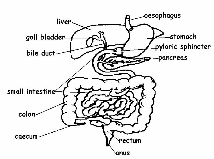
43 colon diagram with labels
Colon Diagram Stock Photos, Pictures & Royalty-Free Images ... Search from Colon Diagram stock photos, pictures and royalty-free images from iStock. Find high-quality stock photos that you won't find anywhere else. Colon Anatomy - Human Body Diagrams - Medical Art Library The large intestine is divided into the cecum, colon, rectum and anal canal.The large intestine begins at the cecum. The ileum (small intestine) ends where it connects to the cecum at the ileocecal junction.. The colon is divided into four parts: the ascending, transverse, descending and sigmoid.The ascending and transverse colon meet at the right hepatic flexure (near the liver).
› stock-photo › female-anatomy-diagramFemale Anatomy Diagram Stock Photos and Images - Alamy Find the perfect female anatomy diagram stock photo. Huge collection, amazing choice, 100+ million high quality, affordable RF and RM images. No need to register, buy now!

Colon diagram with labels
Colon Anatomy (with Small Intestine Label): Image Details 720x602. View. Download. Title: Colon Anatomy (with Small Intestine Label) Description: Drawing shows the cecum, ascending colon, transverse colon, descending colon, sigmoid colon, rectum, and anal canal. Also shown is the small intestine. The cecum connects the small intestine to the colon. Colon (Large Intestine): Anatomy, Function, Structure Sigmoid colon: The S-shaped connection between the last part of the colon and the rectum, located on the bottom left side of the abdomen is called the sigmoid colon. 2 Size and Length This organ is called the large intestine because of the diameter (width) of the intestine; it is much wider than the small intestine, but also much shorter. 40 Colon diagram Vector Images, Colon diagram ... 40 Colon diagram Stock Vector Images, Royalty-free Colon diagram Drawings & Illustrations. VectorMine Crohns disease vector illustration. Labeled diagram with diagnosis. VectorMine Ulcerative colitis vector illustration. Labeled anatomical infographic.
Colon diagram with labels. colon model labeled Flashcards and Study Sets | Quizlet Learn colon model labeled with free interactive flashcards. Choose from 380 different sets of colon model labeled flashcards on Quizlet. Abdomen and digestive system diagrams: normal anatomy | e ... Full labeled anatomical diagrams - Anatomy of the abdomen and digestive system: these general diagrams show the digestive system, with the major human anatomical structures labeled (mouth, tongue, oral cavity, teeth, buccal glands, throat, pharynx, oesophagus, stomach, small intestine, large intestine, liver, gall bladder and pancreas). › enAnatomy, medical imaging and e-learning for ... - IMAIOS IMAIOS and selected third parties, use cookies or similar technologies, in particular for audience measurement. Cookies allow us to analyze and store information such as the characteristics of your device as well as certain personal data (e.g., IP addresses, navigation, usage or geolocation data, unique identifiers). › science › articleSingle-Cell Analyses Inform Mechanisms of Myeloid-Targeted ... Apr 16, 2020 · Single-Cell Analyses Inform Mechanisms of Myeloid-Targeted Therapies in Colon Cancer Author links open overlay panel Lei Zhang 1 6 Ziyi Li 2 6 Katarzyna M. Skrzypczynska 3 6 Qiao Fang 1 Wei Zhang 4 Sarah A. O’Brien 3 Yao He 1 Lynn Wang 3 Qiming Zhang 2 Aeryon Kim 3 Ranran Gao 2 Jessica Orf 3 Tao Wang 2 Deepali Sawant 3 Jiajinlong Kang 2 Dev ...
Histology | Colon This diagram illustrates the 4 basic layers of the colon. The inner pink layer is the mucosa, the yellow layer beneath the mucosa is called the submucosa, while the red layer is the muscular layer (muscularis) and the 4 th layer is called the serosa or adventitia. Courtesy Ashley Davidoff MD 32338 Ultrasound of normal large bowel › tutorials › textword2vec | TensorFlow Core Feb 04, 2022 · All: Speak, speak. First Citizen: You are all resolved rather to die than to famish? All: Resolved. resolved. First Citizen: First, you know Caius Marcius is chief enemy to the people. All: We know't, we know't. First Citizen: Let us kill him, and we'll have corn at our own price. The Colon - Ascending - Transverse - Descending - Sigmoid ... The colon (large intestine) is the distal part of the gastrointestinal tract, extending from the cecum to the anal canal. It receives digested food from the small intestine, from which it absorbs water and electrolytes to form faeces. Anatomically, the colon can be divided into four parts - ascending, transverse, descending and sigmoid. Large intestine with labels for the appendix, cecum ... Large intestine with labels for the appendix, cecum, ascending colon, transverse colon, descending colon, sigmoid colon, rectum, and anus View full-sized image Download Media Please credit each image as: National Institute of Diabetes and Digestive and Kidney Diseases, National Institutes of Health.
The Basics of a Colonoscopy - WebMD Your colon must be completely empty for the colonoscopy to be thorough and complete. To prepare for the procedure, you may have to follow a liquid diet for 1 to 3 days beforehand. A liquid diet ... Colon Diagram Stock Illustrations - 3,263 Colon Diagram ... Human Anatomy. Labeled Diagram. Human colon. Colon - lymphatic drainage. Pathways of lymphatic drainage of the colon. Anatomy of the Colon, Rectum and Anus. Digestive system, large intestine lymphatic. Colonoscopy procedure labeled 3d diagram on white background. Eps 10. Label Digestive System Diagram Printout ... Read the definitions below, then label the digestive system anatomy diagram. anus - the opening at the end of the digestive system from which feces (waste) exits the body. appendix - a small sac located on the cecum. ascending colon - the part of the large intestine that run upwards; it is located after the cecum. Colon: Anatomy, histology, composition, function | Kenhub The colon forms part of the large intestine and extends between the caecum and the rectum. It is about 1.5 meters in length and consists of four parts: ascending transverse descending sigmoid colon You can recognize it easily through several distinct morphological features like semilunar folds and pouches called haustra.
Illustration Picture of Anatomy - Colon The colon is the largest part of the large intestine, extending from the cecum to the rectum. It is 5 feet long and its function is to reabsorb water from digested food and concentrate solid waste material, known as stool. The colon is made of several sections. The ascending colon travels up the right side of the abdomen.
Post a Comment for "43 colon diagram with labels"